
Emerging neuroscientific investigations reveal that electrical stimulation of the vagus nerve—the longest cranial nerve extending from brainstem to abdomen—may represent a paradigm-shifting therapeutic approach for chronic inflammatory conditions, offering mechanistic precision that pharmacological interventions cannot replicate while avoiding systemic side effects characteristic of conventional anti-inflammatory medications.
Chronic inflammation underlies pathophysiology across seemingly disparate disease categories—rheumatoid arthritis, inflammatory bowel disease, cardiovascular disease, neurodegenerative disorders, and metabolic syndrome—creating a public health burden affecting hundreds of millions globally while consuming substantial healthcare resources through lifelong pharmaceutical management that addresses symptoms without resolving underlying dysregulation. The recognition that the vagus nerve mediates a “cholinergic anti-inflammatory pathway” through which the nervous system actively suppresses peripheral inflammation has catalyzed intensive research into vagus nerve stimulation (VNS) as therapeutic modality, with clinical trials demonstrating efficacy across multiple inflammatory conditions while mechanistic studies elucidate the molecular signaling cascades through which electrical nerve activation translates into immune system modulation. This convergence of clinical efficacy and mechanistic understanding positions vagal neuromodulation as potentially transformative intervention that harnesses endogenous regulatory circuits rather than introducing exogenous chemical agents, representing a fundamental reconceptualization of how inflammatory diseases might be treated.
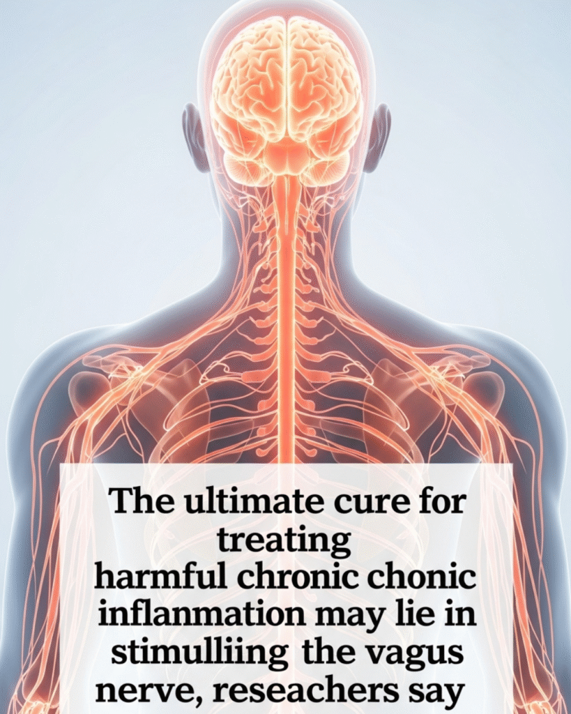
What Is the Vagus Nerve and Why Does It Matter for Inflammation?
The vagus nerve, designated as cranial nerve X in anatomical nomenclature, constitutes the principal component of the parasympathetic nervous system—the branch of autonomic regulation governing “rest-and-digest” physiological states that counterbalance sympathetic “fight-or-flight” activation. Understanding the vagus nerve’s anatomical distribution, functional diversity, and newly discovered immunoregulatory roles provides essential context for comprehending how electrical stimulation might modulate inflammatory processes.
Anatomically, the vagus nerve originates in the medulla oblongata within the brainstem, with cell bodies clustered in the dorsal motor nucleus and nucleus ambiguus. From these origins, vagal fibers descend bilaterally through the neck alongside the carotid arteries, branching extensively to innervate virtually every major organ system: the larynx and pharynx (controlling vocalization and swallowing), the heart (regulating heart rate and contractility), the lungs (modulating bronchial tone and respiratory rate), the gastrointestinal tract from esophagus through transverse colon (controlling motility, secretion, and barrier function), and accessory digestive organs including liver, pancreas, and spleen.
The vagus nerve comprises approximately 80% afferent (sensory) fibers transmitting information from peripheral organs to the brain, with only 20% efferent (motor) fibers carrying commands from brain to periphery. This asymmetry reveals the vagus primarily as a sensory information highway monitoring visceral organ status and conveying this intelligence to central regulatory centers, with the brain using this constant stream of interoceptive data to coordinate appropriate autonomic responses.
The vagal innervation pattern creates anatomical substrate for the nerve’s anti-inflammatory functions. Vagal efferent fibers terminate in ganglia associated with organs including the spleen—the body’s largest lymphoid organ and a major reservoir of immune cells. The discovery that vagal activation could suppress splenic cytokine production emerged from unexpected observations in sepsis research, where electrical vagus nerve stimulation protected experimental animals against lethal endotoxemia through mechanisms independent of the nerve’s previously recognized cardiovascular and respiratory functions.
The molecular mediator of vagal anti-inflammatory effects is acetylcholine—the primary neurotransmitter released by parasympathetic nerve terminals. Acetylcholine binds to nicotinic receptors (specifically the α7 nicotinic acetylcholine receptor subtype) expressed on immune cells including macrophages, dendritic cells, and T lymphocytes. Receptor activation triggers intracellular signaling cascades that suppress pro-inflammatory cytokine production (particularly tumor necrosis factor-alpha, interleukin-1β, and interleukin-6) while potentially enhancing anti-inflammatory mediator release (including interleukin-10). This cholinergic suppression of inflammation operates through multiple mechanisms including inhibition of NF-κB signaling—a master transcriptional regulator controlling inflammatory gene expression—and activation of JAK2/STAT3 pathways that promote anti-inflammatory phenotypes.
The vagus nerve’s anti-inflammatory capacity reflects evolutionary integration of nervous and immune systems, allowing rapid, spatially precise immune modulation that complements slower, more diffuse hormonal regulation. This “inflammatory reflex” enables the brain to detect peripheral inflammation through vagal afferents sensing cytokines and inflammatory mediators, process this information centrally, and deploy vagal efferents to suppress excessive or misdirected immune responses that might otherwise cause tissue damage. The system operates as a negative feedback loop maintaining inflammatory homeostasis, analogous to thermostat-controlled heating systems maintaining temperature stability.
How Does Vagus Nerve Stimulation Suppress Inflammatory Responses?
The mechanistic pathway through which electrical vagus nerve stimulation translates into peripheral immune system modulation involves a complex sequence spanning neural activation, neurotransmitter release, receptor binding, intracellular signaling, and ultimately altered gene expression in immune cells. Elucidating this pathway has required integrating expertise across neuroscience, immunology, molecular biology, and electrophysiology, revealing unexpected interdependencies between systems previously conceived as largely autonomous.
Vagus nerve stimulation typically employs implanted electrode cuffs delivering brief electrical pulses—typically 30 seconds of stimulation at frequencies between 10-30 Hz, pulse widths around 500 microseconds, and intensities below the threshold for aversive sensations or undesired autonomic effects. These parameters were initially optimized for epilepsy treatment (the first approved VNS indication) but have been adapted for anti-inflammatory applications based on empirical testing and emerging mechanistic understanding.
The electrical stimulation activates vagal efferent fibers—the minority population carrying signals from brain to periphery. Action potentials propagate along these myelinated axons at velocities approaching 20 meters per second, reaching peripheral ganglia and synapses within seconds of stimulation initiation. The rapid transmission enables near-instantaneous immune modulation, contrasting sharply with the hours-to-days onset of pharmaceutical anti-inflammatory effects.
At peripheral synapses, arriving action potentials trigger calcium influx that promotes synaptic vesicle fusion and acetylcholine release into the synaptic cleft. The neurotransmitter diffuses across this narrow gap, binding to nicotinic acetylcholine receptors on postsynaptic membranes. For anti-inflammatory effects, the critical targets are α7 nicotinic receptors on macrophages and other immune cells residing in spleen, lymph nodes, intestinal lamina propria, and other lymphoid tissues.
Receptor activation initiates signaling cascades with multiple branches converging on inflammatory gene suppression. The α7 receptor, when bound by acetylcholine, undergoes conformational changes allowing ion flux that alters membrane potential while simultaneously triggering metabotropic signaling through associated proteins. Key pathways include:
JAK2/STAT3 activation: The α7 receptor associates with Janus kinase 2 (JAK2), which phosphorylates signal transducer and activator of transcription 3 (STAT3) upon receptor activation. Phosphorylated STAT3 translocates to the nucleus where it promotes transcription of anti-inflammatory genes including SOCS3 (suppressor of cytokine signaling 3), which inhibits pro-inflammatory signaling pathways through negative feedback.
NF-κB inhibition: Nuclear factor kappa-light-chain-enhancer of activated B cells (NF-κB) represents the master regulator of inflammatory gene expression, controlling transcription of cytokines, chemokines, adhesion molecules, and inflammatory enzymes. Acetylcholine-α7 receptor signaling prevents NF-κB nuclear translocation through mechanisms including prevention of IκB degradation (IκB normally sequesters NF-κB in the cytoplasm), thereby blocking transcriptional activation of inflammatory genes.
MAPK pathway modulation: Mitogen-activated protein kinase (MAPK) cascades including ERK, p38, and JNK pathways transmit inflammatory signals from membrane receptors to nuclear transcription factors. Cholinergic signaling attenuates these pathways, reducing downstream inflammatory mediator production.
Inflammasome regulation: The NLRP3 inflammasome—a multiprotein complex that processes pro-interleukin-1β into its active form—shows reduced activation following vagal stimulation, potentially through mechanisms involving mitochondrial dynamics and reactive oxygen species regulation.
The aggregate effect of these converging pathways is profound suppression of pro-inflammatory cytokine production—particularly TNF-α, IL-1β, IL-6, and IL-18—the mediators driving tissue damage in chronic inflammatory diseases. Simultaneously, anti-inflammatory mediators including IL-10 and TGF-β show enhanced production, shifting the cytokine milieu from pro-inflammatory to regulatory phenotypes that promote resolution rather than perpetuation of inflammation.
Critically, vagal anti-inflammatory effects operate locally within innervated tissues rather than systemically throughout the body, providing spatial precision impossible with pharmaceutical approaches. An oral medication distributes throughout the circulatory system, suppressing immunity globally and potentially compromising infection defense. Vagal stimulation, by contrast, modulates inflammation specifically in innervated lymphoid organs while preserving immune function in non-innervated compartments, theoretically maintaining antimicrobial defenses while suppressing pathological inflammation.
Which Clinical Conditions Might Benefit from Vagal Neuromodulation?
The breadth of diseases involving chronic inflammation as primary or contributory pathophysiology creates extensive potential applications for vagus nerve stimulation, with clinical trials investigating efficacy across diverse conditions that share inflammatory mechanisms despite differing in organ systems affected and traditional diagnostic categorizations.
Rheumatoid Arthritis and Autoimmune Inflammatory Arthritis
Rheumatoid arthritis (RA)—a chronic autoimmune condition causing joint inflammation, pain, swelling, and progressive cartilage and bone destruction—affects approximately 1% of the global population while imposing substantial disability and reduced life expectancy. Current treatments including methotrexate, TNF-α inhibitors, and other immunosuppressive agents provide partial benefit but rarely achieve complete remission while carrying risks of infection and malignancy from systemic immune suppression.
Pilot clinical trials of VNS in RA patients have demonstrated encouraging results. In a small proof-of-concept study, daily vagus nerve stimulation for 12 weeks produced clinically significant improvements in disease activity scores, with some patients achieving remission or low disease activity states. Mechanistically, the intervention reduced serum TNF-α levels—the cytokine targeted by expensive biologic medications—suggesting that electrical neuromodulation might replicate pharmaceutical TNF-α blockade through endogenous mechanisms.
The spatial precision of vagal anti-inflammatory effects may prove particularly advantageous in RA, where suppressing joint inflammation while maintaining systemic antimicrobial immunity would reduce infection risk compared to global immunosuppression. Additionally, the absence of medication-associated hepatotoxicity, nephrotoxicity, or bone marrow suppression potentially expands treatment access to patients intolerant of conventional pharmacotherapy.
Inflammatory Bowel Disease
Crohn’s disease and ulcerative colitis—collectively termed inflammatory bowel disease (IBD)—affect millions worldwide, causing chronic intestinal inflammation, abdominal pain, diarrhea, and increased colorectal cancer risk. The vagus nerve extensively innervates the gastrointestinal tract, providing anatomical substrate for local anti-inflammatory effects within the gut wall where pathology is concentrated.
Preclinical models demonstrate that vagal stimulation reduces intestinal inflammation severity, preserves mucosal barrier integrity, and prevents colitis progression through mechanisms involving both cholinergic anti-inflammatory pathways and modulation of enteric nervous system function. The vagus regulates gut motility, secretion, and barrier function beyond inflammatory control, suggesting that VNS might address multiple pathophysiological dimensions of IBD simultaneously.
Early-phase clinical trials in IBD patients have shown feasibility and preliminary efficacy signals, with some patients experiencing symptom reduction and mucosal healing—the therapeutic goal increasingly recognized as superior to mere symptom palliation. The chronic, relapsing-remitting nature of IBD creates particular need for therapies that can be deployed long-term without cumulative toxicity, a criterion that vagal neuromodulation potentially satisfies.
Cardiovascular Inflammatory Conditions
Atherosclerosis—the arterial disease underlying myocardial infarction and ischemic stroke—is increasingly recognized as inflammatory in character, with immune cells and inflammatory mediators driving plaque formation, progression, and rupture. Additionally, heart failure involves chronic low-grade inflammation contributing to progressive cardiac remodeling and functional decline.
The vagus nerve extensively innervates the heart, regulating rate, contractility, and autonomic balance. Vagal tone—the degree of baseline vagal activity—correlates inversely with cardiovascular mortality, with higher vagal tone predicting better outcomes. VNS has been investigated for heart failure treatment, with some trials showing improved cardiac function and reduced mortality, potentially mediated partially through anti-inflammatory effects that attenuate pathological cardiac remodeling.
The application of VNS to atherosclerosis prevention or treatment remains speculative but mechanistically plausible, given that reducing systemic inflammatory markers (particularly C-reactive protein) through vagal activation might slow plaque progression while stabilizing existing plaques against rupture.
Neuroinflammation and Neurodegenerative Disease
Alzheimer’s disease, Parkinson’s disease, and other neurodegenerative conditions involve neuroinflammation—chronic activation of microglia and astrocytes producing inflammatory mediators that accelerate neuronal death. While the vagus nerve does not directly innervate brain parenchyma (it terminates primarily in brainstem nuclei), vagal stimulation influences brain function through ascending projections and through modulation of peripheral inflammation that affects central nervous system function.
Intriguing preliminary evidence suggests VNS might slow cognitive decline in Alzheimer’s disease or improve motor symptoms in Parkinson’s disease, potentially through reducing peripheral inflammatory signals that exacerbate neuroinflammation or through more direct neuromodulatory effects on brainstem circuits influencing cortical and subcortical function.
Sepsis and Acute Inflammatory Syndromes
While chronic inflammation has been the primary VNS research focus, the most dramatic preclinical results emerged from sepsis models—acute, life-threatening inflammation triggered by infection. Electrical vagal stimulation provided near-complete protection against lethal endotoxemia in animal models, preventing the cytokine storm that causes multi-organ failure and death in septic patients.
Translation to clinical sepsis treatment faces challenges including the acute nature requiring immediate intervention and the heterogeneity of septic presentations. However, the proof-of-principle that neural stimulation can suppress even fulminant inflammatory responses demonstrates the potential power of cholinergic anti-inflammatory pathways when properly activated.
What Technological Approaches Enable Therapeutic Vagus Nerve Stimulation?
Translating mechanistic understanding into clinical therapeutics requires overcoming substantial technological challenges related to electrode design, stimulation parameter optimization, implantation procedures, power delivery, and device longevity. The evolution of VNS technology reflects broader trends in bioelectronic medicine—an emerging field seeking to treat disease through electrical modulation of neural circuits rather than chemical perturbation through pharmaceuticals.
Invasive Implantable Systems
Currently approved VNS devices employ surgically implanted systems analogous to cardiac pacemakers. The core components include:
Electrode lead: A helical cuff electrode wraps around the left cervical vagus nerve (left side is preferred to avoid cardiac effects from right vagal stimulation), providing electrical interface between device and nerve. The electrode design must achieve reliable electrical coupling without causing nerve compression or ischemia that would impair function.
Pulse generator: An implantable pulse generator (IPG) containing battery, stimulation circuitry, and control logic resides in a subcutaneous pocket typically in the left upper chest. The generator delivers programmable electrical pulses to the electrode through an insulated lead tunneled subcutaneously from chest to neck.
External programmer: Clinicians use external telemetry devices to non-invasively program stimulation parameters including frequency, pulse width, intensity, and duty cycle (on-time versus off-time ratios). This programmability allows individualized optimization based on therapeutic response and side effect profiles.
Current-generation devices deliver continuous or intermittent stimulation according to programmed schedules, lacking closed-loop capabilities that would adjust stimulation in real-time based on physiological feedback. Battery life typically ranges from 5-10 years depending on stimulation parameters, after which surgical replacement becomes necessary.
The invasive nature of implantation—requiring general anesthesia and surgical expertise—creates barriers to widespread adoption including cost, infection risk, and patient reluctance. Surgical complications, while uncommon, include hoarseness (from inadvertent recurrent laryngeal nerve injury), dysphagia, infection, and device malfunction requiring replacement.
Non-Invasive Transcutaneous Approaches
To circumvent implantation barriers, researchers have developed transcutaneous VNS (tVNS) delivering electrical stimulation through skin surface electrodes positioned over anatomical locations where the vagus nerve or its branches lie superficially accessible. The two primary targets are:
Auricular branch: The auricular branch of the vagus nerve provides sensory innervation to portions of the external ear, particularly the cymba concha (the depression anterior to the antihelix). Surface electrodes placed in this region can stimulate vagal afferents that project to brainstem nuclei, potentially triggering descending anti-inflammatory efferent activity through reflex arcs.
Cervical transcutaneous stimulation: Electrodes placed over the carotid sheath in the neck can potentially activate the main vagal trunk through transcutaneous electrical nerve stimulation (TENS), though penetration depth and current spread raise questions about stimulation specificity.
Non-invasive approaches offer substantial advantages including eliminating surgical risks, enabling easy therapy discontinuation, and dramatically reducing cost. However, they face challenges in achieving stimulation intensity and specificity comparable to implanted devices, as substantial current must traverse skin and subcutaneous tissue before reaching neural targets, creating discomfort that limits tolerable intensity.
Clinical trials of tVNS for inflammatory conditions have shown mixed results, with some studies reporting efficacy comparable to invasive VNS while others find minimal benefit. These inconsistencies likely reflect variations in stimulation parameters, electrode positioning, patient selection, and the inherent challenge of achieving adequate vagal activation through skin surface electrodes. Ongoing research seeks to optimize tVNS protocols and identify biomarkers predicting which patients will respond to non-invasive approaches.
Closed-Loop Bioelectronic Systems
The frontier of VNS technology involves closed-loop systems that sense physiological signals and adjust stimulation in real-time to maintain therapeutic targets—analogous to how insulin pumps with continuous glucose monitoring adjust insulin delivery based on measured blood glucose.
For anti-inflammatory applications, closed-loop VNS would ideally sense inflammatory markers (cytokine levels, immune cell activation states) and modulate vagal stimulation to maintain inflammation within homeostatic ranges while minimizing stimulation time and intensity. Implementation faces challenges in developing implantable biosensors capable of reliably measuring relevant biomarkers continuously over years.
Alternative closed-loop approaches might use surrogate signals more easily measured—heart rate variability (which reflects vagal tone), blood pressure, or other autonomic parameters—as proxies for inflammatory status, adjusting stimulation to maintain these metrics within target ranges. While indirect, such approaches might provide sufficient regulation to improve efficacy while reducing side effects compared to open-loop continuous stimulation.
The integration of machine learning algorithms that learn individual patient responses could further optimize therapy, identifying patterns in physiological signals that predict inflammatory flares and preemptively modulating vagal activity to prevent symptomatic episodes.
How Do Clinical Trial Results Support Vagal Anti-Inflammatory Therapy?
The translation of preclinical mechanistic insights into clinical benefit requires rigorous evaluation through controlled trials balancing internal validity (experimental control minimizing bias) against external validity (generalizability to real-world patient populations and clinical settings). The VNS-for-inflammation trial landscape reveals promising signals tempered by the methodological challenges inherent to device trials and the heterogeneity of inflammatory disease presentations.
Rheumatoid Arthritis Clinical Evidence
The SetPoint Medical RESET-RA trial, published in 2016, represented a landmark proof-of-concept study demonstrating VNS efficacy in drug-resistant RA. Seventeen patients who had failed multiple conventional disease-modifying antirheumatic drugs (DMARDs) received implanted VNS devices delivering intermittent stimulation (60 seconds four times daily) over 84 days.
Results showed clinically significant improvements on standardized RA assessment scales (DAS28—Disease Activity Score in 28 joints), with several patients achieving low disease activity or remission states previously unattainable despite aggressive pharmacotherapy. Importantly, responders showed reductions in serum TNF-α levels correlating with clinical improvement, providing human evidence that the mechanism identified in animal models translates to patients.
The study’s open-label design (no sham stimulation control group) limits definitive efficacy conclusions, as placebo effects prove substantial in RA trials. However, the magnitude and durability of responses, combined with mechanistic biomarker changes, suggest genuine therapeutic effects beyond expectancy. Subsequent larger, controlled trials are underway to definitively establish efficacy while identifying predictors of response that might guide patient selection.
Inflammatory Bowel Disease Preliminary Data
Small pilot studies have investigated VNS in Crohn’s disease patients, generally showing feasibility and preliminary efficacy signals. A study by Bonaz and colleagues implanted VNS devices in seven Crohn’s disease patients, delivering stimulation over 6 months. Six patients showed clinical improvement with reduced symptom scores, while five showed biological improvement (reduced inflammatory markers and/or mucosal healing on endoscopy).
These encouraging results must be interpreted cautiously given the small sample size and open-label design. IBD shows substantial placebo response rates (improvements despite receiving inactive treatments), and the natural history includes spontaneous remissions that might coincide with trial participation. Nonetheless, the observation that vagal stimulation can induce mucosal healing—not merely symptom reduction—suggests genuine disease modification rather than symptomatic palliation.
Sepsis and Critical Care Applications
While chronic inflammatory conditions have dominated VNS research, the original discovery emerged from sepsis models where vagal activation prevented lethal inflammation. Translation to human sepsis treatment faces unique challenges: sepsis requires acute intervention without time for device implantation, patient heterogeneity is extreme, and standard-of-care has evolved to include targeted therapies addressing specific infectious causes.
Nonetheless, the potential to rapidly suppress cytokine storms through electrical stimulation remains compelling. Recent pilot studies have explored feasibility of emergency VNS placement in septic patients, with some showing reduced inflammatory markers and organ dysfunction. The very small sample sizes preclude efficacy conclusions, but technical feasibility has been demonstrated, establishing foundation for larger trials.
Methodological Challenges and Future Directions
VNS trials face methodological challenges complicating efficacy assessment:
Sham control difficulties: Ideal placebo-controlled trials include sham devices providing no stimulation, but participants often detect whether they receive active stimulation through sensations (throat tightness, voice changes) or cardiovascular effects (heart rate changes), compromising blinding. Novel sham protocols attempt to address this through sub-threshold stimulation or stimulation of non-therapeutic nerves, but perfect blinding remains elusive.
Parameter optimization uncertainty: The vast parameter space (frequency, pulse width, intensity, duty cycle) means current stimulation protocols reflect empirical optimization rather than comprehensive systematic evaluation. Individual patients might respond optimally to different parameters, but identifying personalized optimal settings requires adaptive trial designs not yet widely implemented.
Long-term efficacy and safety: Most trials span months, insufficient for assessing chronic treatment over years or decades that inflammatory disease management requires. Long-term complications—nerve damage from chronic electrode contact, device failures, infection—must be characterized through extended follow-up.
Patient selection and response prediction: Inflammatory diseases show substantial patient heterogeneity, with individuals varying in disease mechanisms, severity, and treatment responses. Identifying biomarkers predicting VNS response would enable precision medicine approaches directing therapy to likely responders, but such predictive markers remain largely unidentified.
What Are the Safety Considerations and Potential Side Effects?
Any therapeutic intervention entails benefit-risk trade-offs requiring comprehensive safety characterization to inform clinical decision-making. Vagus nerve stimulation, while generally well-tolerated, produces predictable side effects related to stimulation of vagal efferent and afferent fibers innervating structures beyond intended inflammatory targets, while rare serious complications require consideration particularly when evaluating therapy for non-life-threatening conditions.
Common Stimulation-Related Side Effects
The most frequent adverse effects directly result from electrical activation of vagal fibers innervating the larynx, pharynx, and cardiovascular system:
Voice changes and hoarseness: Stimulation activates recurrent laryngeal nerve branches controlling vocal cord abduction, producing hoarseness, voice alteration, or vocal fatigue during stimulation periods. Most patients adapt with effects diminishing over weeks as accommodation occurs, while parameter adjustments (reducing intensity or pulse width) often mitigate symptoms while maintaining therapeutic benefit.
Cough and throat tightness: Pharyngeal and laryngeal sensations including tingling, tightness, or cough occur in substantial patient fractions, typically coinciding with stimulation pulses. These effects generally prove tolerable and decrease with accommodation.
Dysphagia: Swallowing difficulties occasionally occur when stimulation disrupts coordinated esophageal motility, though severe dysphagia remains rare.
Cardiovascular effects: The vagus extensively innervates the heart, regulating heart rate and atrioventricular conduction. Stimulation can reduce heart rate (bradycardia) or increase heart rate variability. While clinically significant cardiac effects are uncommon with left vagal stimulation at typical parameters, cardiac monitoring during parameter adjustment is prudent, particularly in patients with pre-existing cardiac conduction abnormalities.
Headache and dizziness: Non-specific symptoms including headache, dizziness, and nausea occur in some patients, potentially related to brainstem stimulation through afferent pathways or to autonomic effects.
The majority of these effects prove mild to moderate in severity, dose-dependent (intensity-related), and reversible with stimulation cessation or parameter adjustment. Importantly, VNS does not produce the immunosuppression-related infections or malignancies associated with conventional immunosuppressive medications, representing a fundamental safety advantage for chronic use.
Surgical and Device-Related Complications
Implantable VNS requires surgical procedures carrying inherent risks:
Infection: Surgical site infections or device-related infections occur in approximately 1-5% of cases, sometimes necessitating device explantation and antibiotic therapy. Infection risk reduction employs standard surgical sterile technique, prophylactic antibiotics, and careful wound management.
Nerve injury: Though rare, inadvertent nerve damage during lead placement can cause permanent voice changes or swallowing difficulties if the recurrent laryngeal nerve sustains injury.
Lead migration or fracture: Electrode leads can migrate from optimal positioning or fracture from repetitive neck movement, requiring revision surgery.
Device malfunction: Electronic devices occasionally fail through battery depletion, circuit malfunction, or component degradation, necessitating replacement.
These surgical risks must be weighed against disease severity and alternative treatment options. For severe, refractory inflammatory conditions causing substantial disability despite maximal medical therapy, the procedural risks may prove acceptable, while less severe conditions might warrant prioritizing non-invasive approaches or awaiting further technology development.
Theoretical Long-Term Concerns
Chronic vagal stimulation might theoretically produce unforeseen consequences through sustained modulation of autonomic balance:
Parasympathetic overdominance: Excessive vagal activation could theoretically produce bradycardia, reduced cardiac contractility, or gastrointestinal dysmotility, though clinical experience to date has not demonstrated problematic parasympathetic excess at therapeutic stimulation parameters.
Immune suppression: While targeted anti-inflammatory effects represent the therapeutic goal, excessive or prolonged suppression might theoretically increase infection susceptibility. However, the preserved immune function in non-innervated compartments combined with the absence of increased infections in clinical trials suggests this theoretical concern may not materialize clinically.
Accommodation and tolerance: Chronic stimulation might induce neural or receptor changes reducing therapeutic efficacy over time, analogous to tolerance development with pharmaceutical agents. Long-term efficacy data will determine whether this occurs.
Which Future Directions Might Enhance Vagal Anti-Inflammatory Therapy?
The current state of vagal neuromodulation for inflammation represents early phases of technological and therapeutic evolution, with numerous research trajectories promising to enhance efficacy, reduce invasiveness, and expand applications through advances spanning neuroscience, engineering, and clinical medicine.
Targeted Fiber-Selective Stimulation
The vagus nerve comprises multiple fiber types serving different functions—motor fibers controlling vocalization and swallowing, cardiovascular regulatory fibers, gastrointestinal control fibers, and inflammatory reflex pathways. Current stimulation activates these fiber populations relatively non-selectively, creating side effects from unintended fiber activation.
Advanced electrode designs employing spatially-distributed contacts with independent control could theoretically achieve fiber-selective stimulation—activating anti-inflammatory pathways while avoiding voice and swallowing fibers. This requires detailed understanding of vagal fiber topography (which fiber types occupy which positions within the nerve bundle) combined with computational modeling of current spread patterns from different electrode configurations.
Alternatively, stimulation parameter optimization might exploit differences in fiber activation thresholds—different fiber populations require different current intensities or frequencies for activation—allowing selective activation through carefully tuned parameters. Preliminary preclinical work suggests this approach’s feasibility, but clinical translation requires systematic parameter space exploration.
Molecular and Genetic Enhancement of Vagal Function
Rather than merely stimulating existing vagal pathways, future approaches might enhance the pathways themselves through molecular interventions:
Optogenetic approaches: Genetic modification to express light-sensitive ion channels (opsins) in specific vagal neuron populations would enable optical rather than electrical stimulation, potentially allowing unprecedented specificity. While currently confined to research contexts, advancing gene therapy delivery and wireless optogenetic stimulation could eventually enable clinical applications.
Pharmacological modulation of cholinergic signaling: Rather than electrical stimulation, medications enhancing acetylcholine availability at immune cell synapses or potentiating α7 receptor signaling could theoretically replicate anti-inflammatory effects. Acetylcholinesterase inhibitors (blocking acetylcholine breakdown) or α7 receptor-specific agonists represent potential pharmaceutical alternatives to neuromodulation, though achieving therapeutic immune modulation without central nervous system side effects presents challenges.
Vagal tone enhancement through lifestyle: Emerging evidence suggests that certain interventions increase baseline vagal tone—meditation, slow breathing exercises, cold exposure, specific dietary patterns—potentially providing non-invasive methods to enhance endogenous anti-inflammatory regulation. While unlikely to match targeted electrical stimulation for severe inflammatory conditions, such approaches might reduce disease burden at population levels or serve as adjunctive therapies.
Integration with Digital Health and AI
The convergence of implantable neuromodulation devices with digital health technologies creates opportunities for enhanced monitoring and therapy optimization:
Continuous inflammatory monitoring: Implantable biosensors measuring inflammatory markers in real-time could enable closed-loop systems adjusting stimulation based on measured inflammation levels rather than fixed schedules.
Machine learning optimization: Algorithms analyzing patterns in physiological signals, patient-reported outcomes, and inflammatory biomarkers could identify optimal stimulation parameters for individual patients, accelerating the trial-and-error process currently required for parameter optimization.
Remote monitoring and telehealth integration: Connected devices transmitting data to healthcare providers enable proactive intervention for device issues or disease flares, while reducing clinic visit burdens.
Expansion to Additional Inflammatory Conditions
As mechanistic understanding deepens and clinical experience accumulates, VNS applications will likely expand beyond currently investigated conditions to other inflammatory diseases sharing common pathophysiology. Potential future targets include:
Metabolic syndrome and obesity: Chronic low-grade inflammation contributes to insulin resistance, atherosclerosis, and metabolic dysfunction. Vagal stimulation might address multiple metabolic syndrome components simultaneously through anti-inflammatory effects combined with metabolic regulation.
Chronic pain syndromes: Many chronic pain conditions involve neuroinflammation contributing to pain persistence and sensitization. Vagal anti-inflammatory effects might reduce pain through inflammation suppression while direct analgesic effects from ascending vagal pathways provide additional benefit.
Psychiatric conditions: Depression and anxiety show associations with elevated inflammatory markers, and emerging evidence suggests anti-inflammatory interventions might provide antidepressant or anxiolytic effects in inflammatory subgroups. VNS is already approved for treatment-resistant depression through mechanisms incompletely understood but potentially involving anti-inflammatory pathways.
Conclusion: Toward Precision Neuromodulation of Inflammatory Disease
The recognition that the nervous system actively regulates peripheral inflammation through the vagus nerve represents a paradigm expansion in immunology, revealing intimate bidirectional communication between neural and immune systems that prior conceptual frameworks largely overlooked. This discovery creates therapeutic opportunities to harness endogenous anti-inflammatory circuitry through targeted electrical stimulation, potentially providing disease-modifying treatments for chronic inflammatory conditions that conventional pharmacology addresses inadequately while causing substantial side effects.
Clinical trial evidence, while preliminary, demonstrates proof-of-principle that vagus nerve stimulation can suppress inflammation and improve clinical outcomes in conditions including rheumatoid arthritis and inflammatory bowel disease. The mechanistic pathway from electrical stimulation through acetylcholine release to immune cell α7 receptor activation and inflammatory gene suppression has been elucidated with impressive detail, providing biological plausibility and molecular targets for therapy optimization.
Challenges remain in technological development, parameter optimization, patient selection, and long-term safety characterization. Current invasive implantable systems create access barriers while non-invasive approaches have not yet achieved reliability comparable to implantation. The ideal stimulation parameters likely vary between individuals and conditions, requiring personalized approaches not yet systematically implemented.
The evolution toward precision neuromodulation—employing targeted, closed-loop, fiber-selective stimulation guided by continuous inflammatory monitoring and machine learning optimization—promises to enhance efficacy while reducing side effects. Such systems would transform vagal anti-inflammatory therapy from the relatively crude, open-loop approaches of current generation devices to sophisticated interventions rivaling pharmaceutical precision while avoiding systemic medication drawbacks.
For the millions suffering from chronic inflammatory diseases inadequately controlled by conventional treatments, vagus nerve stimulation represents a fundamentally different therapeutic paradigm—activating the body’s own regulatory systems rather than introducing exogenous chemicals that inevitably produce unintended effects. While not yet ready for routine clinical deployment outside specialized centers and research contexts, the trajectory suggests that vagal neuromodulation will increasingly complement or even replace pharmaceutical anti-inflammatory therapy across the coming decades, fulfilling the promise of bioelectronic medicine to treat disease through targeted electrical modulation of neural circuits.
Important Disclaimer: This article presents scientific and medical information for educational purposes and should not be construed as medical advice. Vagus nerve stimulation for inflammatory conditions remains investigational for most applications, with approval status varying by jurisdiction and indication. Individuals with inflammatory diseases should consult qualified healthcare providers to discuss treatment options appropriate for their specific circumstances. The