Meta Description: Learn key findings in chest radiography and how to recognize six common conditions. A clear guide to interpreting chest X-rays in clinical pathology.
Chest radiography (chest X-ray) is one of the most widely used diagnostic tools in medicine. It provides a fast, non-invasive way to evaluate the lungs, heart, and chest structures. For clinicians and students, learning to recognize key findings in chest radiography is essential for identifying common and potentially serious conditions early.
In this guide, we’ll explore six frequently encountered chest pathologies and their typical radiographic features to help you build a strong foundation in interpretation.
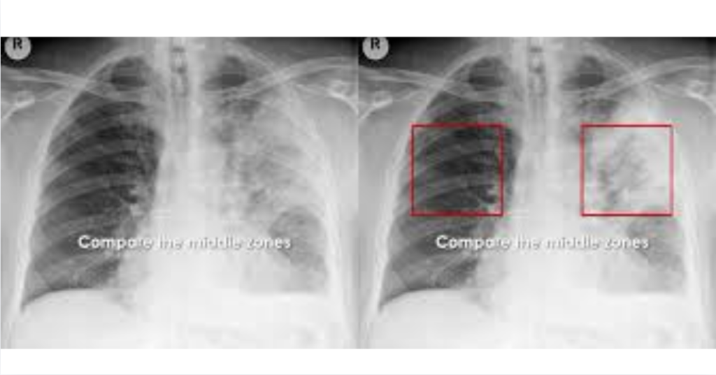
What Is Chest Radiography?
Chest radiography uses low-dose X-rays to create images of the structures inside the thorax.
What it evaluates
- Lungs
- Heart size and shape
- Pleura (lining of lungs)
- Diaphragm
- Mediastinum
Why it is important
- Fast and widely available
- First-line imaging tool
- Helps detect life-threatening conditions
Featured Snippet: What is chest radiography used for?
Chest radiography is used to evaluate the lungs, heart, and chest structures, helping detect infections, fluid buildup, heart enlargement, and other thoracic conditions.
1. Pneumonia
Pneumonia is a lung infection that causes alveolar inflammation.
Key radiographic findings
- Localized or lobar consolidation
- Air bronchograms (air-filled bronchi visible)
- Increased lung opacity
Clinical correlation
- Fever
- Cough
- Chest pain
2. Pleural Effusion
Pleural effusion is fluid accumulation in the pleural space.
Key radiographic findings
- Blunted costophrenic angles
- Meniscus sign (curved fluid line)
- White-out in severe cases
Common causes
- Heart failure
- Infection
- Malignancy
Featured Snippet: What does pleural effusion look like on X-ray?
Pleural effusion appears as fluid accumulation in the lung base with blunted costophrenic angles and a characteristic meniscus-shaped opacity.
3. Chronic Obstructive Pulmonary Disease
COPD is a chronic lung disease causing airflow limitation.
Key radiographic findings
- Hyperinflated lungs
- Flattened diaphragm
- Increased retrosternal airspace
- Reduced vascular markings
Clinical correlation
- Chronic cough
- Shortness of breath
- History of smoking
4. Pneumothorax
Pneumothorax is air in the pleural space causing lung collapse.
Key radiographic findings
- Visible pleural line
- Absence of lung markings beyond the line
- Collapsed lung edge
Emergency signs
- Tension pneumothorax may shift mediastinum
- Medical emergency
Featured Snippet: What is pneumothorax on chest X-ray?
Pneumothorax appears as a visible pleural line with absent lung markings beyond it, indicating air in the pleural space and partial or complete lung collapse.
5. Cardiomegaly (Heart Enlargement)
Cardiomegaly refers to an enlarged heart shadow.
Key radiographic findings
- Increased cardiothoracic ratio (>50%)
- Enlarged cardiac silhouette
- Possible associated pulmonary congestion
Common causes
- Heart failure
- Hypertension
- Cardiomyopathy
6. Pulmonary Edema
Pulmonary edema is fluid accumulation in lung tissue.
Key radiographic findings
- Bilateral “bat-wing” opacities
- Kerley B lines (interstitial edema)
- Enlarged heart (if cardiac cause)
Clinical correlation
- Shortness of breath
- Orthopnea
- Crackles on auscultation
Featured Snippet: What does pulmonary edema look like on X-ray?
Pulmonary edema appears as bilateral fluffy opacities, often in a “bat-wing” pattern, along with signs of fluid overload such as Kerley B lines.
How to Systematically Read a Chest X-Ray
A structured approach improves accuracy.
Step 1: Check image quality
- Positioning
- Exposure
- Rotation
Step 2: Airway and mediastinum
- Trachea position
- Mediastinal width
Step 3: Heart size
- Cardiothoracic ratio
- Shape and borders
Step 4: Lung fields
- Opacities
- Hyperinflation
- Asymmetry
Step 5: Diaphragm and pleura
- Costophrenic angles
- Fluid or air presence
Step 6: Bones and soft tissues
- Rib fractures
- Abnormal shadows
Common Pitfalls in Chest Radiography Interpretation
1. Poor inspiration
- Can mimic disease
2. Rotation errors
- Distorts heart and mediastinum
3. Overlapping shadows
- Can hide pathology
Featured Snippet: What are common chest X-ray findings?
Common chest X-ray findings include pneumonia (consolidation), pleural effusion (fluid buildup), COPD (hyperinflation), pneumothorax (air in pleural space), cardiomegaly (enlarged heart), and pulmonary edema (fluid in lungs).
Clinical Importance of Chest Radiography
Why it is essential
- Rapid diagnosis in emergencies
- Monitoring chronic diseases
- Guiding treatment decisions
Limitations
- Limited soft tissue detail
- May require CT for confirmation
Internal Linking Opportunities
This topic connects well with:
- Respiratory diseases and lung function
- Cardiovascular imaging and heart health
- Emergency medicine and trauma care
- Infectious diseases like pneumonia
- Radiology interpretation basics
Conclusion
Chest radiography remains a cornerstone of medical imaging, offering quick and valuable insights into a wide range of thoracic conditions. Recognizing key findings in common diseases such as pneumonia, pleural effusion, COPD, pneumothorax, cardiomegaly, and pulmonary edema is essential for accurate diagnosis and timely care.
With a structured approach and consistent practice, chest X-ray interpretation becomes a powerful clinical skill that enhances patient outcomes and diagnostic confidence.
Important Disclaimer: This article is for educational purposes only and should not replace professional medical training or clinical judgment. For health-related concerns, consult qualified healthcare professionals.