Meta Description: Learn the 12 cranial nerves, their key functions, and a simple anterior view guide to understanding how they control sensation, movement, and vital reflexes.
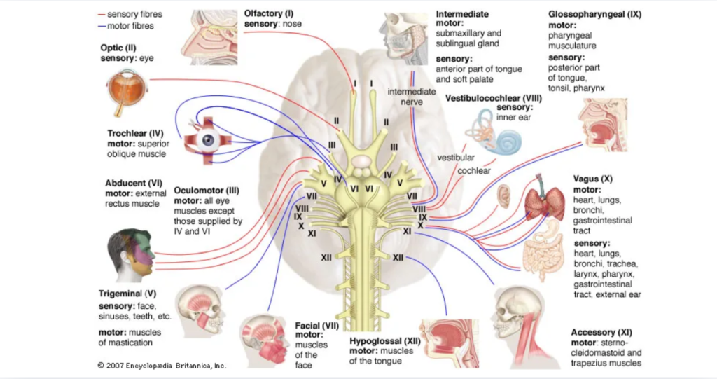
The cranial nerves are a set of 12 paired nerves that emerge directly from the brain, primarily the brainstem. They are responsible for transmitting sensory, motor, and autonomic signals between the brain and structures of the head, neck, and some internal organs.
Understanding them from an anterior (front) perspective helps visualize how they distribute through the face, eyes, nose, mouth, and neck.
Featured Snippet: What are cranial nerves and what do they do?
Cranial nerves are 12 pairs of nerves that originate from the brain and control functions such as smell, vision, facial movement, hearing, balance, swallowing, and autonomic control of internal organs.
The 12 Cranial Nerves (Anterior Functional Overview)
I – Olfactory nerve
Olfactory nerve
- Function: Smell
- Pathway: Nose → olfactory bulb → brain
II – Optic nerve
Optic nerve
- Function: Vision
- Connects retina to the brain
III – Oculomotor nerve
Oculomotor nerve
- Moves most eye muscles
- Controls pupil size and lens adjustment
IV – Trochlear nerve
Trochlear nerve
- Helps eye move downward and inward
V – Trigeminal nerve
Trigeminal nerve
- Sensation of face
- Jaw movement (chewing muscles)
VI – Abducens nerve
Abducens nerve
- Moves eye outward (abduction)
VII – Facial nerve
Facial nerve
- Facial expressions
- Taste (front 2/3 of tongue)
- Tear and salivary glands
VIII – Vestibulocochlear nerve
Vestibulocochlear nerve
- Hearing
- Balance
IX – Glossopharyngeal nerve
Glossopharyngeal nerve
- Taste (posterior tongue)
- Swallowing
- Saliva production
X – Vagus nerve
Vagus nerve
- Heart rate regulation
- Digestion
- Voice and swallowing
XI – Accessory nerve
Accessory nerve
- Shoulder movement
- Head rotation
XII – Hypoglossal nerve
Hypoglossal nerve
- Tongue movement
- Speech and swallowing
Functional Grouping of Cranial Nerves
Sensory nerves
- I (Olfactory)
- II (Optic)
- VIII (Vestibulocochlear)
Motor nerves
- III, IV, VI (eye movement)
- XI (neck/shoulder)
- XII (tongue)
Mixed nerves
- V (Trigeminal)
- VII (Facial)
- IX (Glossopharyngeal)
- X (Vagus)
Anterior Perspective: Why It Matters
From a front view, cranial nerves are mainly associated with:
- Facial sensation (V)
- Facial expression (VII)
- Eye movement (III, IV, VI)
- Smell and vision (I, II)
- Swallowing and speech (IX, X, XII)
This perspective is especially useful in clinical neurology examinations.
Clinical Importance
Cranial nerve testing helps evaluate:
- Brainstem function
- Stroke effects
- Nerve injury
- Neurological diseases
Related concept
Neurological examination
Common Disorders Affecting Cranial Nerves
- Bell’s palsy (facial nerve dysfunction)
- Trigeminal neuralgia (facial pain)
- Vagus nerve dysfunction (voice/swallowing issues)
- Optic nerve damage (vision loss)
Related condition
Bell’s palsy
Common Myths
Myth: Cranial nerves only affect the head
They also control vital internal organ functions, especially the vagus nerve.
Myth: Damage always causes complete loss of function
Partial nerve damage may produce mild or localized symptoms.
Myth: All cranial nerve problems are permanent
Some conditions, like inflammation or compression, can be reversible.
Internal Linking Opportunities
This topic connects well with:
- Brainstem function
- Neurological examination
- Stroke and nerve damage
- Sensory and motor systems
- Clinical anatomy education
Conclusion
The 12 cranial nerves form a critical communication network between the brain and the head, neck, and internal organs. Understanding their functions from an anterior perspective makes it easier to visualize how they control sensation, movement, and essential physiological processes.
A clear grasp of cranial nerve anatomy is essential for interpreting neurological symptoms and supporting clinical assessment.
Important Disclaimer: This article is for educational purposes only and should not replace professional medical advice. Neurological symptoms should be evaluated by a qualified healthcare professional.